 English
 Français
 Español
 Deutsch
 Italiano
 العربية



Tel
:+86-755-86961139
E-Mail
:sales@vivo-light.com
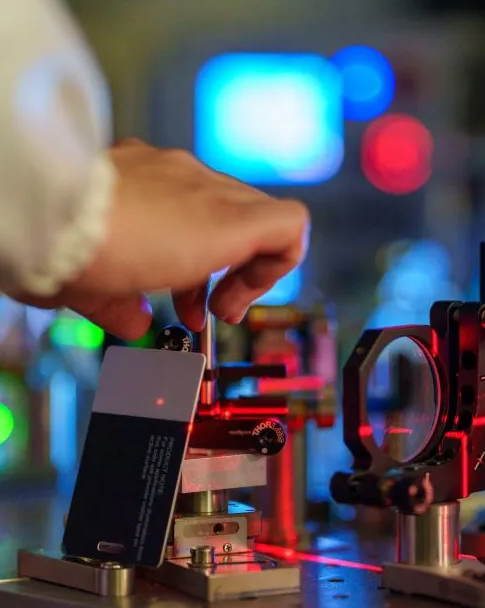

Ein Infrarot-Adernfinder Das Gerät sendet Nahinfrarotlicht in das Hautgewebe aus. Dort absorbiert das desoxygenierte Hämoglobin in den subkutanen Blutgefäßen das Licht, während es von den umliegenden Strukturen reflektiert wird. Dieser optische Mechanismus projiziert eine visuelle Karte direkt auf die Haut. Der Venenfinder macht das blinde Abtasten bei schwierigen Venenpunktionen überflüssig. Sie können sofort auch versteckte Venen identifizieren, die durch bloßes Tasten nicht ertastet werden können. In komplexen Krankenhausbehandlungen haben 59,3 % der Patienten Schwierigkeiten beim Venenzugang. Der Einsatz eines Infrarot-Venenfinders ersetzt das Rätselraten durch eine klare Echtzeitdarstellung. Sie finden sofort geeignete Gefäßwege und erhöhen so die Erfolgsrate beim ersten Punktionsversuch. Diese nicht-invasive visuelle Hilfestellung erhöht die Patientensicherheit bei jeder routinemäßigen Blutentnahme.
Ein Infrarot-Venenfinder erkennt subkutane Blutgefäße durch gezielte Lichtabsorption. Hält man das tragbare Gerät über die Haut, beleuchtet es das darunterliegende Gewebe mit Licht im Spektrum von 700 bis 900 nm. Menschliches Gewebe bleibt in diesem Nahinfrarotbereich transparent, sodass Photonen 10 bis 15 mm tief in die subkutanen Schichten eindringen können. Desoxygeniertes Hämoglobin in den roten Blutkörperchen absorbiert Nahinfrarotlicht mit Absorptionsmaxima bei etwa 758 nm und 929 nm. Umliegende subkutane Strukturen reflektieren diese Lichtwellen zurück zum Sensor, wodurch messbare Absorptionsunterschiede entstehen. Verschiedene menschliche Gewebe weisen bei Lichteinwirkung unterschiedliche mittlere Streukoeffizienten auf.
Gewebetyp | Mittlerer Streukoeffizient (μs) |
Haut | 46,0 |
Knochen | 22,9 |
Gehirn | 24.2 |
Andere Weichteile | 18.9 |
Fettgewebe | 18.4 |
Brust | 16.8 |
Quelle: Henderson et al., Frontiers in Neurology, 2024, „Kann Infrarotlicht wirklich das bewirken, was wir ihm zuschreiben? Prinzipien, Praktiken und Grenzen der Infrarotlichtpenetration.“
Dieser physikalische Kontrast erzeugt ein scharfes optisches Signal. Fettgewebe und Weichteile reflektieren einfallende Strahlen aufgrund ihrer geringeren Lichtabsorption. Hautgewebe streut Licht stark, sodass die Eindringtiefe mit zunehmender Tiefe rasch abnimmt. Studien zur Eindringtiefe von 820 nm Infrarotlicht zeigen, dass die Transmission mit zunehmender Hautdicke stark abnimmt; in bestimmten experimentellen Hautmodellen ist in 3 mm Tiefe praktisch kein Licht mehr messbar. Die Nahinfrarot-Technologie zielt auf diese optimale obere Hautschicht ab. Mit einem Handgerät lassen sich verborgene Strukturen sofort lokalisieren. Diese nicht-invasive Venenvisualisierungstechnologie erstellt klare visuelle Zielkarten basierend auf anatomischen Unterschieden.
Quelle: Frontiers in Neurology, 2024
Sobald die Spezialkamera reflektierte Lichtmuster erfasst hat, verarbeitet eine integrierte Software die visuellen Daten umgehend. Die Nahinfrarot-Bildgebung arbeitet in Bereichen mit geringer Melaninabsorption und ermöglicht so eine effektive Erkennung bei unterschiedlichen Hauttönen. Die Reflexionsdifferenz-Bildgebung berechnet die Intensitätsunterschiede zwischen Gewebereflexionen und Hämoglobinabsorption. Kantenschärfungsalgorithmen entfernen Hintergrundrauschen und schärfen Strukturkonturen. Spezielle Tiefenvorhersagemodelle schätzen die Venentiefe anhand der Streuintensitätsmuster. Die digitale Signalverarbeitung filtert eingehende Signale innerhalb von Millisekunden und gewährleistet so die sofortige Einsatzbereitschaft.
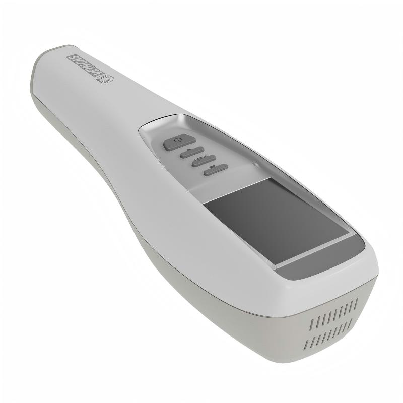
Nach der Verarbeitung des visuellen Signals zeigt das System eine Echtzeit-Venenprojektion auf der Hautoberfläche des Patienten an. tragbares Infrarot-Adernsuchgerät Visuelle Unterstützung bei der Gefäßdarstellung kann die Abhängigkeit von Erfahrungswerten verringern. Ein Handgerät emittiert Infrarotlicht, um optimale Einstichstellen hervorzuheben. Ein moderner Venen-Visierer verbessert periphere Zugangsverfahren, indem er blindes Tasten überflüssig macht. Dieses fortschrittliche Gerät stellt unterirdische Gefäße millimetergenau dar. Kliniker verlassen sich auf den Venen-Visierer, um bei jeder Blutentnahme einen präzisen Zugang zu gewährleisten. So erzielen Sie konstanten Erfolg beim Legen von intravenösen Zugängen, selbst bei anspruchsvollen pädiatrischen und erwachsenen Patienten.
Sie stehen täglich vor der Herausforderung, Venen zu punktieren, wenn Patienten mit geschwollenem Gewebe, dunkler Hautpigmentierung oder fragilen Blutgefäßen vorstellig werden. Moderne Venenvisualisierungstechnologien verbessern die Arbeitsabläufe im klinischen Alltag. Standardmäßige Tastuntersuchungen versagen oft bei ödematösen oder dehydrierten Patienten, da die Flüssigkeit das Zielgefäß verdeckt. Ein tragbares Venenvisualisierungsgerät macht unterirdische Gefäße durch das oberflächliche Gewebe hindurch deutlich sichtbar.
Bei Patienten mit schwierigem Venenzugang, wie z. B. Kindern, älteren Menschen oder dunkelhäutigen Personen, verbessern Venenfinder die Sichtbarkeit und den Erfolg des Eingriffs.
Bei Patienten mit hohem BMI werden moderne Venenfinder eingesetzt, um subkutane Strukturen zu lokalisieren. Fettgewebe bildet eine physische Barriere, die jede periphere Vene unter Fettschichten verbirgt. Ein Infrarot-Venenfinder projiziert helle visuelle Signale direkt auf die Haut, um die Nadelführung zu erleichtern. Bei Patienten mit einem BMI von 30 oder höher ist die Wahrscheinlichkeit, mit Hilfe der Lichtprojektion beim ersten Versuch eine Vene zu finden, etwa 3,5-mal höher.
Quelle: Basadonna G. et al., University of Massachusetts Medical School, klinische Studie mit AccuVein AV400 bei adipösen Patienten.
Moderne Venensuchgeräte steigern die Effizienz von Eingriffen bei Patienten mit Adipositas permagna. Bei Patienten mit einem BMI von 40 oder höher erhöhte sich die Erfolgsrate nach zwei Versuchen von ca. 50 % auf 96 %. Allerdings müssen die optischen Grenzen beachtet werden. Ein tragbares Handgerät erkennt subkutane Gefäße bis zu einer maximalen Tiefe.
Mithilfe eines tragbaren Venensuchgeräts lassen sich oberflächliche Venen bis zu einer Tiefe von 10 mm darstellen. Nahinfrarotlicht kann diese Grenze nicht überschreiten. Liegt eine tiefer liegende Vene mehr als 10 mm unter dichtem Fettgewebe, muss stattdessen eine Ultraschalluntersuchung durchgeführt werden. Der Einsatz eines geeigneten Venensuchgeräts optimiert den intravenösen Zugang bei Routineeingriffen und trägt zur Einhaltung hoher Standards in modernen Gesundheitseinrichtungen bei.
Visuelle Gefäßdarstellungsgeräte können im medizinischen Umfeld bedenkenlos eingesetzt werden. Das in modernen Infrarot-Venenbildgebungsgeräten verwendete Nahinfrarotlicht zählt zu den nichtionisierenden Strahlen. Eine in J Biophotonics (2023) veröffentlichte Studie bestätigte, dass Nahinfrarotlicht weder die DNA schädigt noch Mutationen in menschlichen Fibroblastenzellen auslöst. Die Hersteller entwickeln diese Geräte gemäß den strengen Richtlinien der FDA-Zulassung und den Qualitätsstandards der ISO 13485. Gerätenormen wie IEC 60601-2-57 gewährleisten optimale Kontrollen optischer Gefahren im Routinebetrieb.
Diese Geräte nutzen sicheres, nicht-invasives Licht. Sie berühren weder die Haut noch verursachen sie Schmerzen.
Ein optisches Venenfinder für die Projektion Durch die Erfassung von Lichtabsorptionsunterschieden zwischen den Gefäßen werden die darunterliegenden Gefäßstrukturen deutlich sichtbar. Sauerstoffarmes Blut in den Venen absorbiert spezifische Wellenlängen, während die umgebenden Strukturen diese reflektieren. Zielvenenverläufe lassen sich so leicht visuell erkennen:
1. Das Gerät sendet Infrarotlicht aus, das die Hautoberfläche durchdringt.
2. Das Hämoglobin im Blut absorbiert dieses Infrarotlicht, wodurch das Gerät das Gefäßmuster unter der Haut erkennen kann.
3. Das aufgenommene Gefäßbild wird von der Gerätesoftware verarbeitet.
4. Das verarbeitete Bild wird in Echtzeit auf die Haut des Patienten projiziert und liefert so eine klare visuelle Darstellung der Venen ohne Hautpenetration.
Ein tragbares Handgerät ermöglicht es medizinischem Fachpersonal, optimale Venenpunkte sofort zu identifizieren. Diese nicht-invasive visuelle Darstellung hilft, versehentliche Arterienpunktionen zu vermeiden und das Risiko schmerzhafter Komplikationen wie Hämatombildung oder Blutverlust bei routinemäßigen peripheren Venenpunktionen deutlich zu reduzieren. So erzielen Sie höchste klinische Präzision bei gleichzeitig maximaler Sicherheit in allen Bereichen.
Ein Infrarot-Venensucher visualisiert unterirdische Gefäße durch selektive Lichtabsorption. So lassen sich verborgene Gefäße sofort sichtbar machen und die Effizienz bei schwierigen Eingriffen steigern. Klinische Studien ergaben folgende Ergebnisse: Bei Kindern erhöhte der Einsatz eines Venensuchers die Erfolgsrate beim ersten Versuch von 47,4 % auf 91,7 %. Die durchschnittliche Anzahl der Punktionsversuche sank von 2,23 auf 1,08.
Quelle: PubMed-indexierte randomisierte kontrollierte Studie mit 129 pädiatrischen Patienten. Erhöht die Verwendung eines Venenvisualisierungsgeräts zur Platzierung peripherer Venenkatheter die Erfolgsrate bei pädiatrischen Patienten?
Die Integration eines fortschrittlichen Venenfinders mit Projektion oder eines Infrarot-Venenvisualisators in die Arbeitsabläufe im Gesundheitswesen kann das Risiko einer blinden Punktion verringern. Sie erhalten sofortige Orientierung und sichern gleichzeitig den peripheren Venenzugang bei komplexen Patientenfällen. In einer Umfrage unter Einrichtungen, die Nahinfrarot-Venenvisualisierungstechnologie einsetzen, berichteten 88 % der Befragten von einer höheren Patientenzufriedenheit.
Quelle: Journal of Infusion Nursing, 2023.
Ein tragbares Venen-Darstellungsgerät erkennt subkutane Gefäße bis zu einer Tiefe von 10 mm. Nahinfrarotlicht durchdringt die oberen Hautschichten und hebt den Gefäßverlauf deutlich hervor.
Nein. Diese nicht-invasiven Geräte verwenden harmloses Nahinfrarotlicht. Sie emittieren keine ionisierende Strahlung, sodass sie von medizinischem Fachpersonal bei Routineeingriffen bedenkenlos eingesetzt werden können.
Ein Handgerät projiziert eine visuelle Echtzeitkarte auf die Haut. So lassen sich sofort ideale Punktionspunkte lokalisieren, wodurch schmerzhafte Nadelstiche bei schwierigen Patienten reduziert werden.
Extrem tief liegende Zielgefäße mit einem Durchmesser von mehr als 10 mm erfordern alternative Methoden wie Ultraschall. Optische Beleuchtung kann jedoch die meisten oberflächlichen Zielgefäßverläufe deutlich darstellen.
Eine Nachricht hinterlassen
Scannen zu WhatsApp :


 IPv6 NETZWERK UNTERSTÜTZT